Small Cells, Big Pictures
Former Biological Imaging Facility director Steve Ruzin helped expand UC Berkeley’s microscopy capabilities over a period of more than three decades.
When Steve Ruzin, PhD ’84 Botany, started working at UC Berkeley in 1989 as manager of the Center for Plant Developmental Biology, the fledgling facility was still finding its footing.
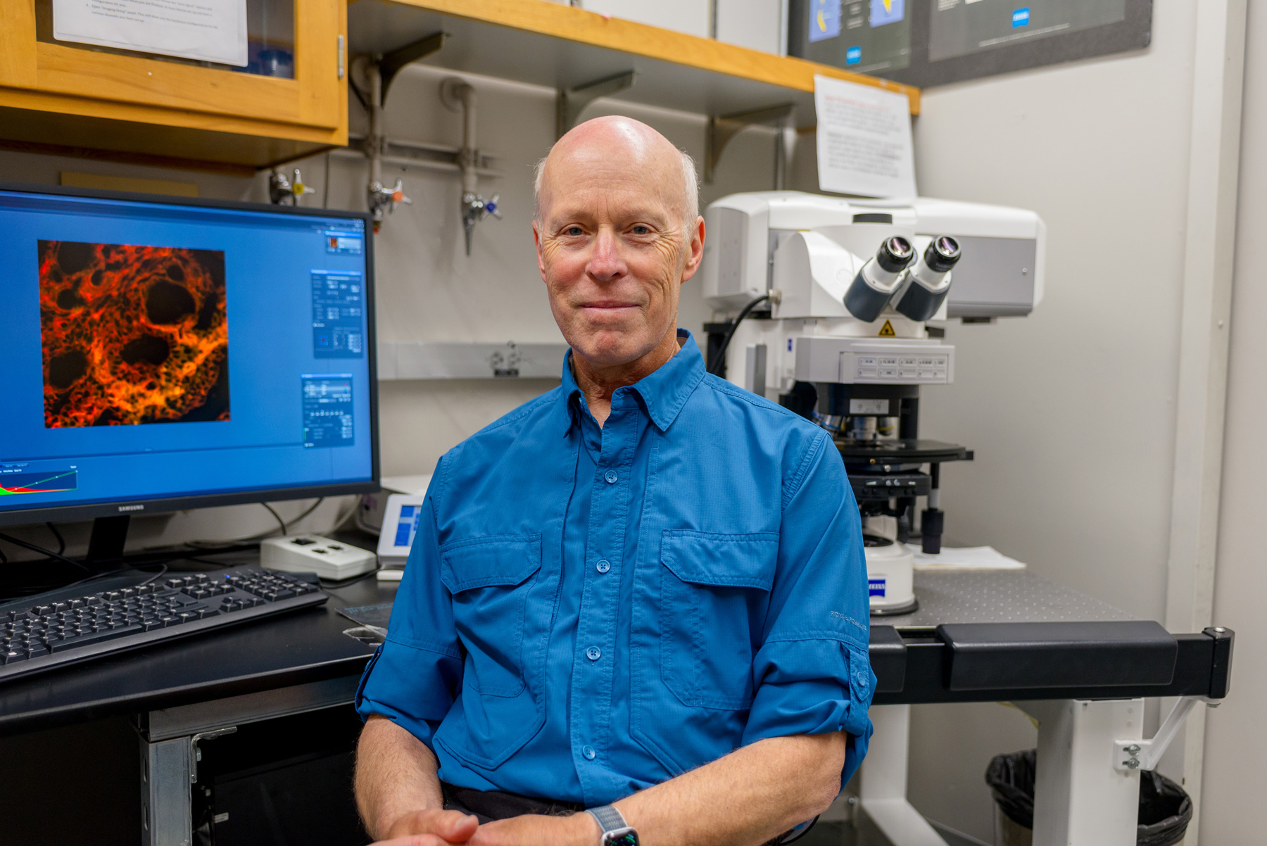
Steve Ruzin in the Biological Imaging Facility. Photo by Mathew Burciaga.
Funded by a National Science Foundation (NSF) award to Professor Mike Freeling in the Department of Plant and Microbial Biology (PMB), the center’s original purpose was to utilize modern and classical techniques in genetics and cell biology to deepen understanding of the development of a variety of specialized cells in corn and other plants. The grant would support two technicians and up to five graduate students, a seminar program, and research funds for faculty affiliated with the Center.
As the field of developmental biology advanced and equipment capabilities evolved, Ruzin predicted that high-magnification microscopes would be useful for researchers across campus. So in 1991, Ruzin used $200,000 from their initial NSF grant to purchase a Sarastro Phoibos 1000, the first commercial laser scanning confocal microscope that was capable of producing a three-dimensional visualization of a sample. “That was probably the first confocal microscope at UC Berkeley,” he said. “I don’t think anyone in the NSF Center had heard of it before.”
As the Center acquired more equipment and increased its user base, Ruzin approached College of Natural Resources leadership with a proposal to transform it into a dedicated microscopy core facility. The proposal was accepted, and in 1998 Ruzin was named director of the Biological Imaging Facility (BIF), one of four core laboratories on campus that offer expertise, instruction, and instrumentation in microscopy for research. He hired Denise Schichnes, PhD ’97 Plant and Microbial Biology, as a lab scientist to help run the facility.
“Microscope technology has changed so much over the years,” said Ruzin, who worked with Schichnes and other faculty labs to secure more than $2.5 million in grant funding for new equipment from the NSF, the National Institutes of Health (NIH), and the Berkeley Faculty Research Fund.

Now, the BIF is capable of high-magnification and high-speed microscopy, examining living cells using fluorescent probes, and imaging through microscope-mounted digital cameras. More than 5,000 scientific publications have benefited from the BIF’s equipment, according to Ruzin’s estimates, and the Facility has hosted upwards of 4,000 users from across the university and elsewhere. Stanford University and UC San Francisco have used the BIF’s equipment to advance their research, as have Bay Area biotech companies and even public radio and television station KQED.
Ruzin retired from full-time duties in 2022 but remains active with the BIF, which—now under the direction of Schichnes—continues to offer cutting-edge tools for researchers. “The Facility just submitted an NIH proposal for a microscope that is very sensitive and really fast,” he adds. “We brought six primary investigators together and showed that we can gather very dim fluorescent observations of cells on this machine—something we can't do anywhere else.”
Evidence of Ruzin’s impact on campus also extends beyond the walls of Koshland Hall. He wrote the first website for PMB in HTML during the Netscape-era, taught undergraduate and graduate-level courses, and has authored textbooks on plant microtechnique and light microscopy. As an emeritus faculty member, Ruzin continues to curate The Golub Collection—which consists of over 160 antique microscopes donated by Orville Golub, PhD ’44 Microbiology—and serves as a guide on tours organized by the Cal Alumni Association.
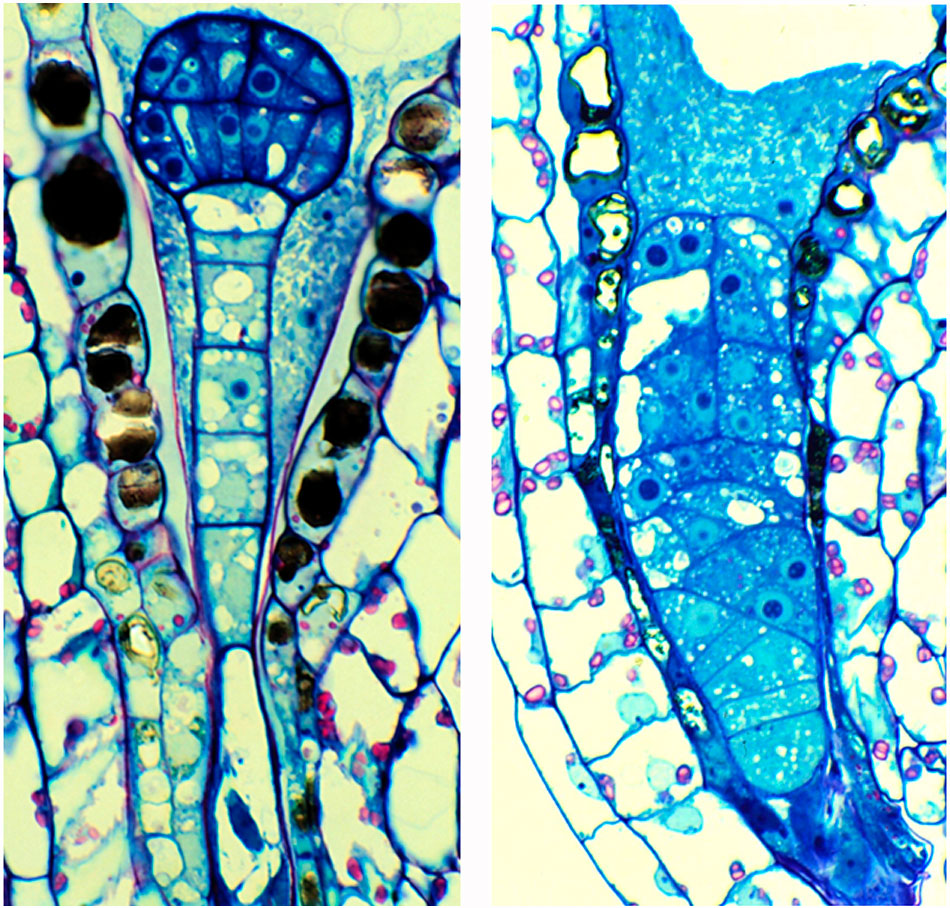
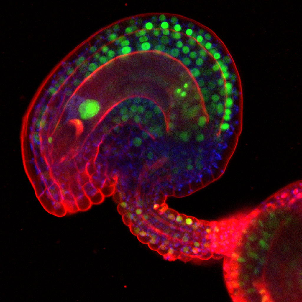
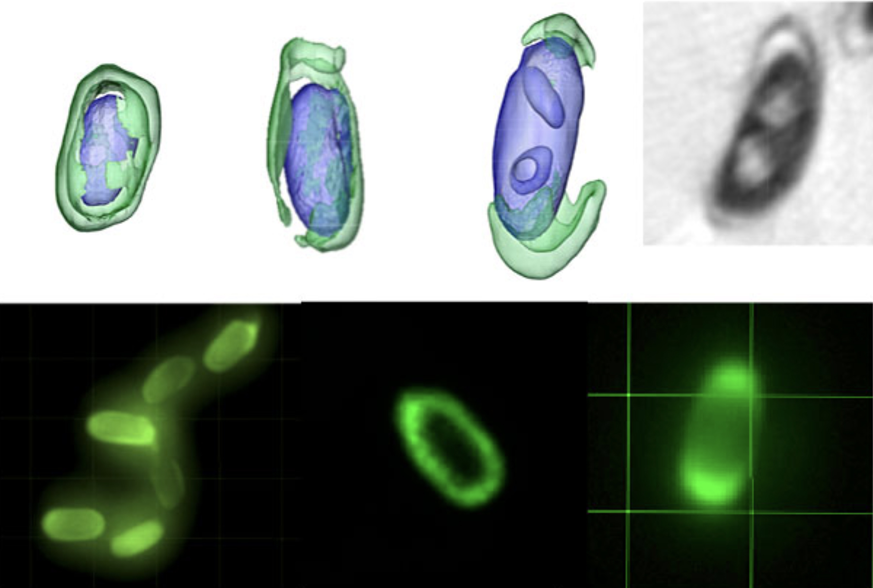
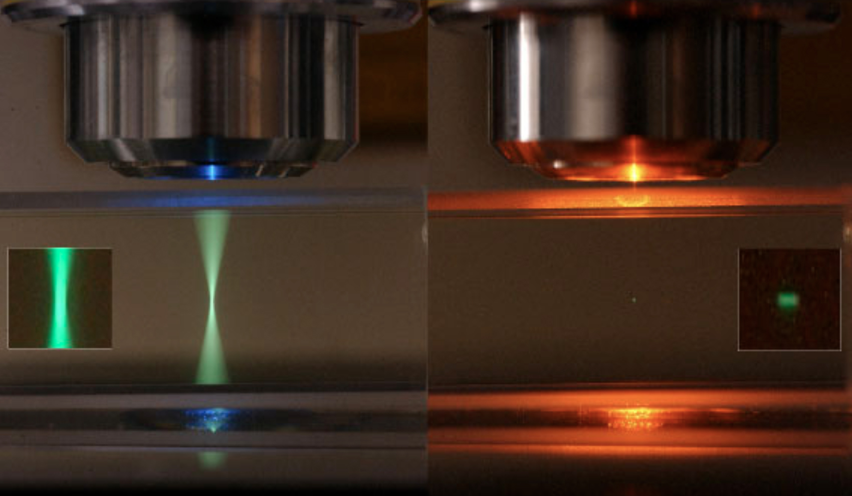
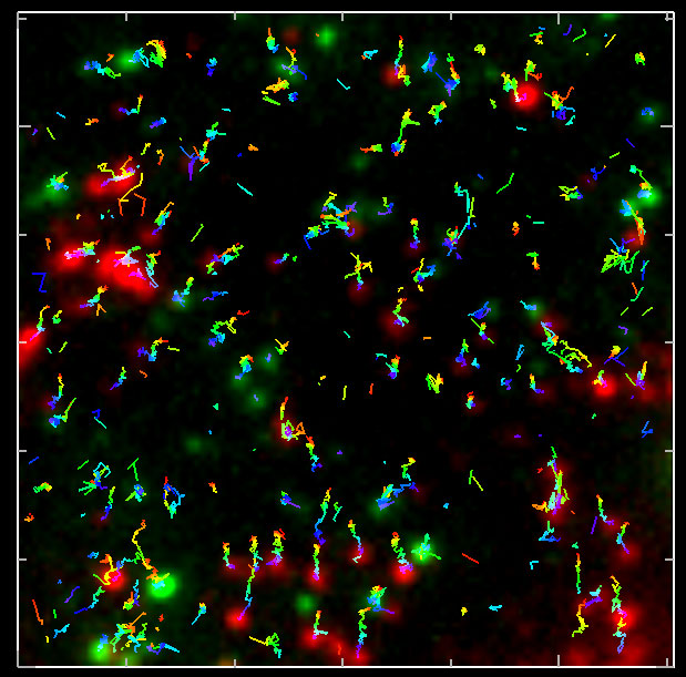
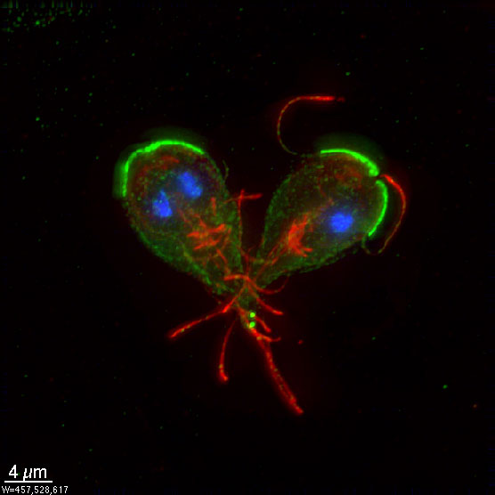
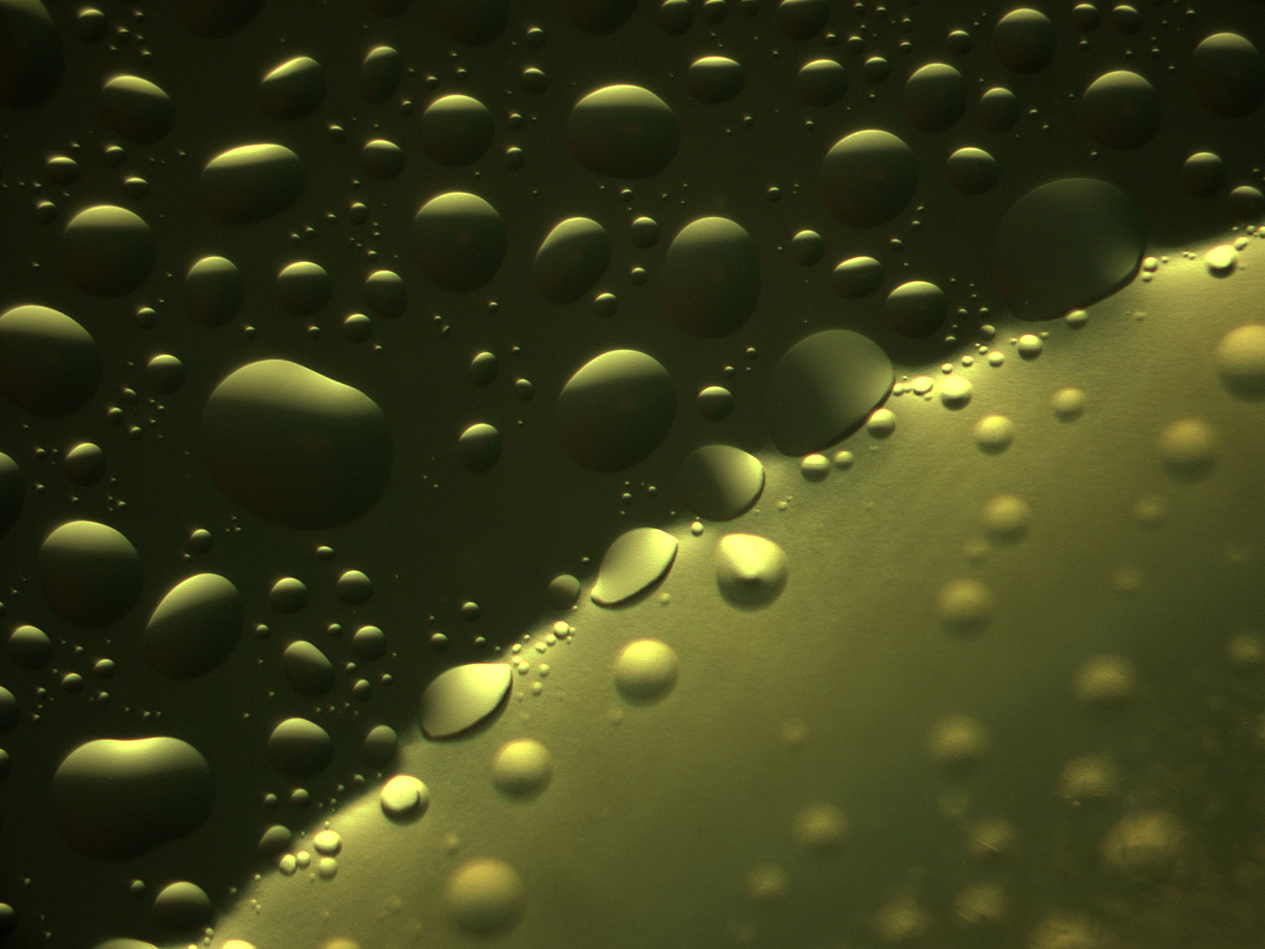
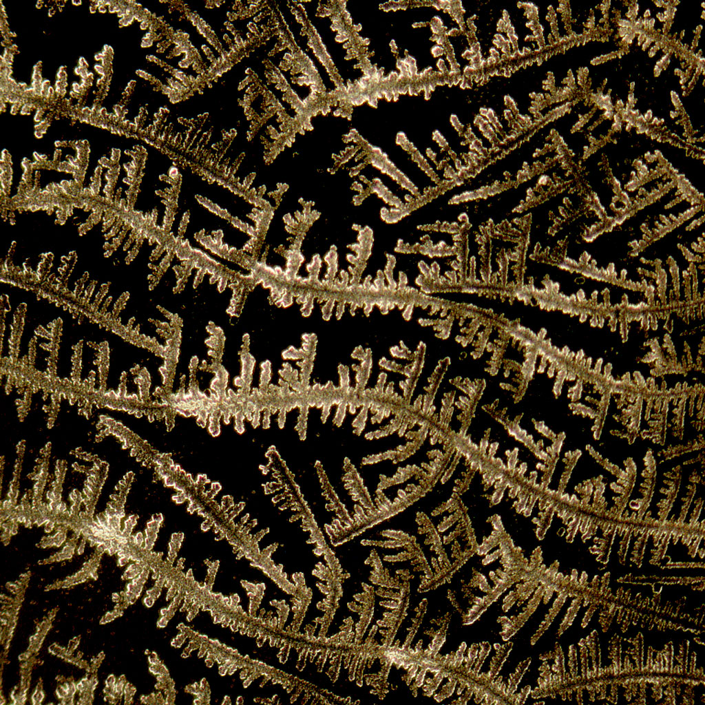
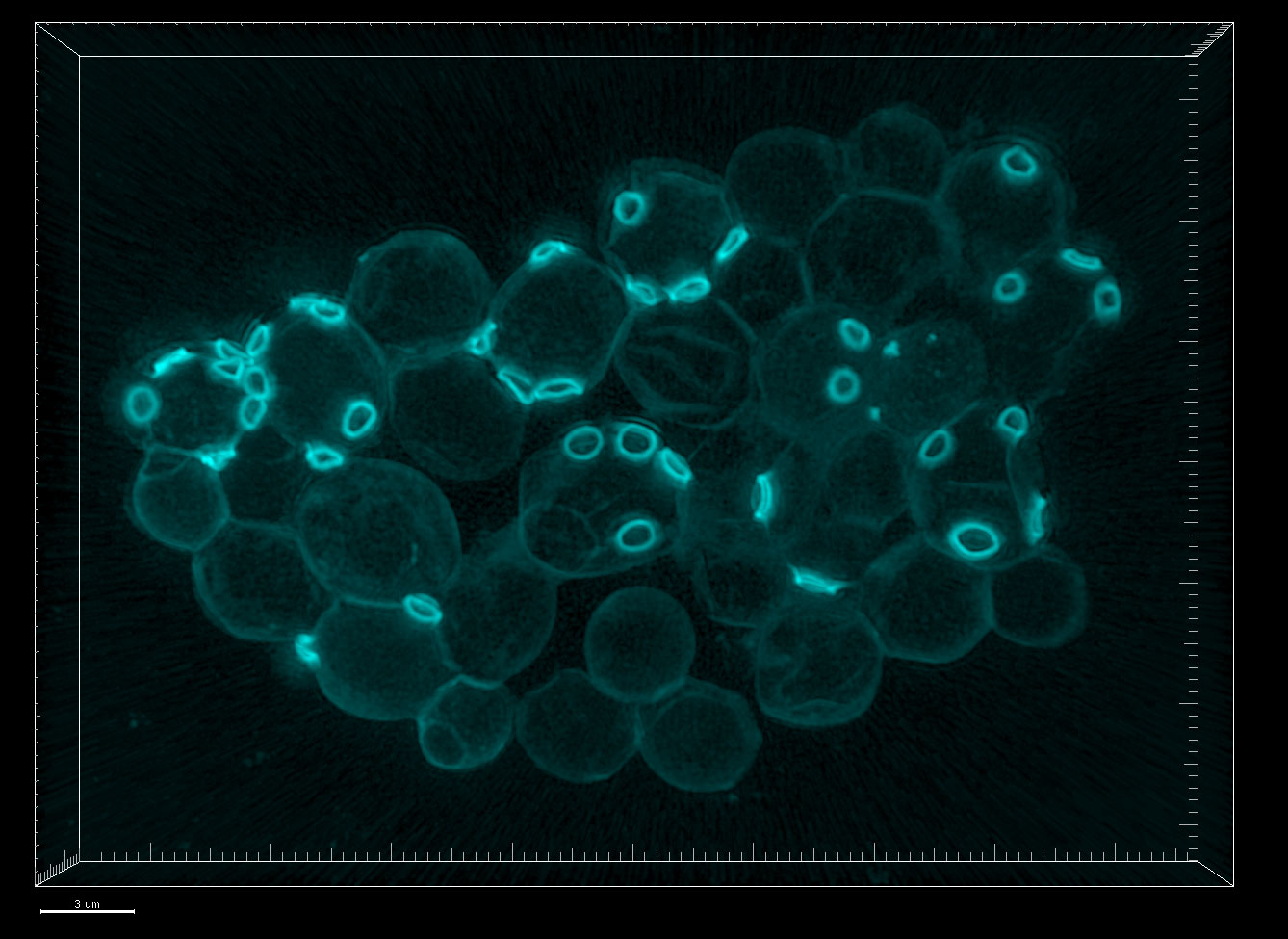
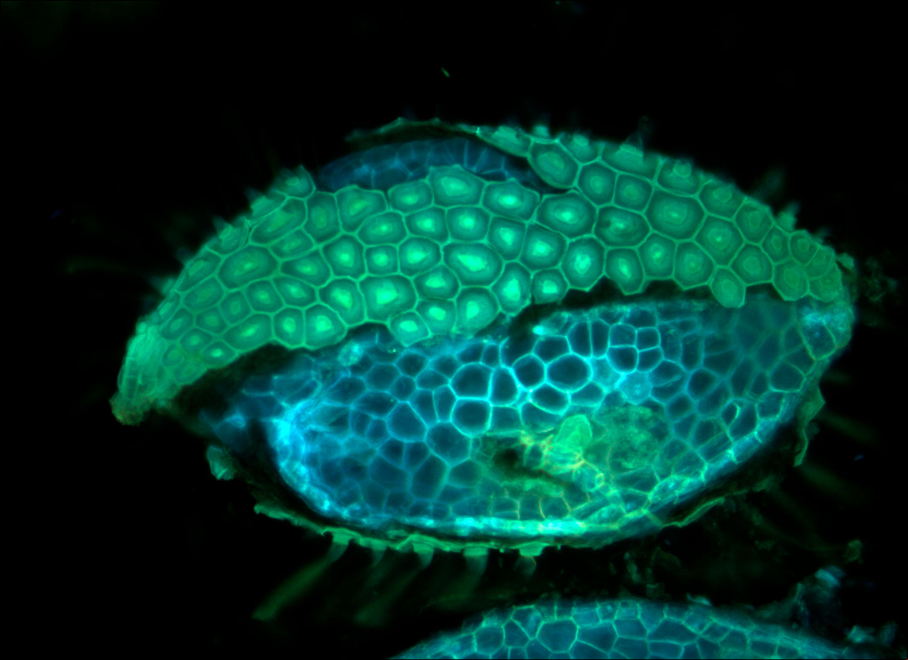
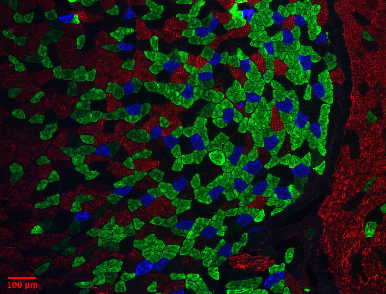
To showcase the beauty of the microscopic world, we asked Ruzin to select a handful of images from the BIF’s long-running “Image of the Month” competition. The images below were all submitted by the Facility’s users and represent a small slice of the fluorescent, three-dimensional, and light microscopy capabilities they offer. Since 2002, Ruzin has personally awarded a box of See’s Chocolates to the winners, and each winning image was featured on the Facility’s homepage for a month.